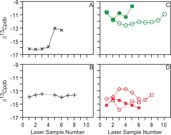
Fig 4. Laser ablation stable carbon isotope data for specimens analyzed in the study.
A, hominin canine AT-825. B, hominin incisor AT-146. C, Cervus elaphus samples: open circles, molar ATA88-TGIIIa- -F21-43; closed circles, lower third molar ATA04-TD10-J21-234. D, Ursus deningeri samples: open squares, left lower first molar SH97-U14-137-Arcillas; closed squares, lower canine SH02-R17-Brecha; diamond, left lower first molar SH02-R/S-16/17-Arcillas. Symbols are larger than the precision (<0.3‰) for each scan.

