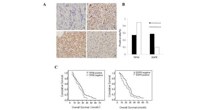
Figure 1.
Expressions of Tat-interacting protein (TIP)30 and epidermal growth factor receptor (EGFR) in glioma and normal brain tissue samples. (A) (a) Negative expression of TIP30 in glioma tissue samples. (b) Positive expression of TIP30 in normal tissue samples. (c) Strong positive expression of EGFR in glioma tissue samples. (d) Weak positive expression of epidermal growth factor receptor (EGFR) in normal brain tissue samples. (Magnification, ×400) (B) Protein expression of TIP30 and EGFR in glioma and normal brain tissue samples (P<0.05). (C) Median overall survival (mOS) of patients with TIP30 and EGFR-positive and TIP30 and EGFR-negative expression. (Left panel) Patients with TIP30-positive expression exhibited longer mOS times, as compared with patients with TIP30-negative expression (P=0.019), and (right panel) patients with EGFR-positive expression exhibited shorter mOS times, as compared with patients with EGFR-negative expression (P=0.021).

