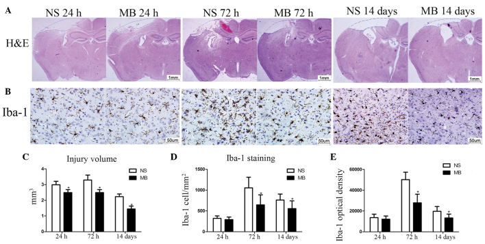
Figure 2.
MB reduces cerebral lesion volume and microglial activation following TBI. (A) Representative images of HE-stained coronal sections 24 h, 72 h and 14 days post-TBI (Scale bar=1 mm). (B) Representative images of Iba-1-stained coronal sections 24 h, 72 h and 14 days post-TBI (Scale bar=50 µm). (C) Quantitative analysis of injury volume revealed a significant decrease in loss of tissue following treatment with MB (n=7; *P<0.05). (D and E) Quantitative analysis demonstrated reduced Iba-1-positive cell numbers and fluorescence intensity following treatment with MB. (n=6; *P<0.05). The data are presented as the mean ± standard deviation. MB, methylene blue; TBI, traumatic brain injury; NS, normal saline; HE, hematoxylin and eosin.

