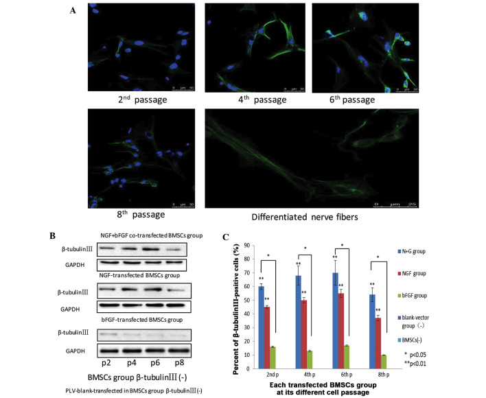
Figure 5.
(A) Nerve growth factor (NGF) and basic fibroblast growth factor (bFGF) co-transfected bone marrow mesenchymal stem cells (BMSCs) at passages 2, 4, 6, and 8 express β-tubulin III, as detected by the presence of green fluorescent dye. Scale bar=50 mm. Neuron-like extensions and inter-connections of NGF and bFGF were depicted. Nerve fibers scale bar=25 mm. All images were obtained with a laser scanning confocal microscope (Zeiss LSM 510). (B) The expression of β-tubulin III in the various groups of differentiated cells at passages 2, 4, 6, and 8. (C) Flow cytometric analysis of β-tubulin III expression in the three groups at passages 2, 4, 6, and 8. Data are presented as the mean ± standard deviation (n=3). *P<0.05, **P<0.01.

