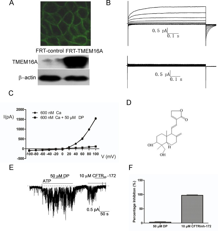
Fig 1. Identification of a small molecule inhibitor (DP) of human TMEM16A.
A, FRT cells stably transfected with human TMEM16A and GFP showing green membrane fluorescence (above) and TMEM16A protein by western blotting (below). B, Examples of whole-cell currents recorded from FRT-TMEM16A cells at a holding potential of 0 mV, followed by pulsing voltages between ±100 mV in steps of 20 mV in the absence (above) or presence of 50 μM DP (below). TMEM16A CaCC currents were elicited by 600 nM of free calcium in pipette solution. C, Current/voltage (I/V) plot of the mean currents at the middle of each voltage pulse. D, Chemical structure of DP. E, CFTR Cl- current trace recorded from CFTR-expressing FRT cells. CFTR Cl- current was elicited by 1 mM ATP, followed by the addition of DP and CFTRinh-172. The dashed line represents zero current. F, The bars represent the percentage inhibition of DP (n = 6) and CFTRinh-172 (n = 4) on CFTR Cl- current.

