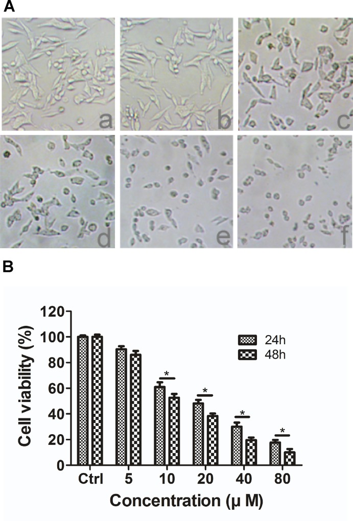
Fig 3. The effect of DP on the morphological characteristics and the viability of SW620 cells.
A, Morphological changes of SW620 cells observed under phase-contrast microscopy (100 × magnification) after treating cells with (a) control, (b) 5 μM, (c) 10 μM, (d) 20 μM, (e) 40 μM or (f) 80 μM of DP for 24 h. B, SW620 cells were treated with various concentrations (0, 5, 10, 20 40 and 80 μM) of DP for 24 or 48 h, as described in the methods. Cell viability was measured using the MTT assay. The results are expressed as the mean ± S.D. (n = 3); * p < 0.05.

