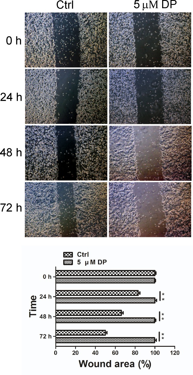
Fig 4. Effects of DP on SW620 cell motility.
Migration of SW620 cells was assessed by a wound-healing assay in the presence of 5 μM DP, compared to the control group. Representative images of wound closure were taken at 0, 24, 48 and 72 h after injury under 40 × magnification (above). Bar graphs of wound area are shown (below). Values are the means ± SD; n = 3; ** p < 0.01. Ctrl, control.

