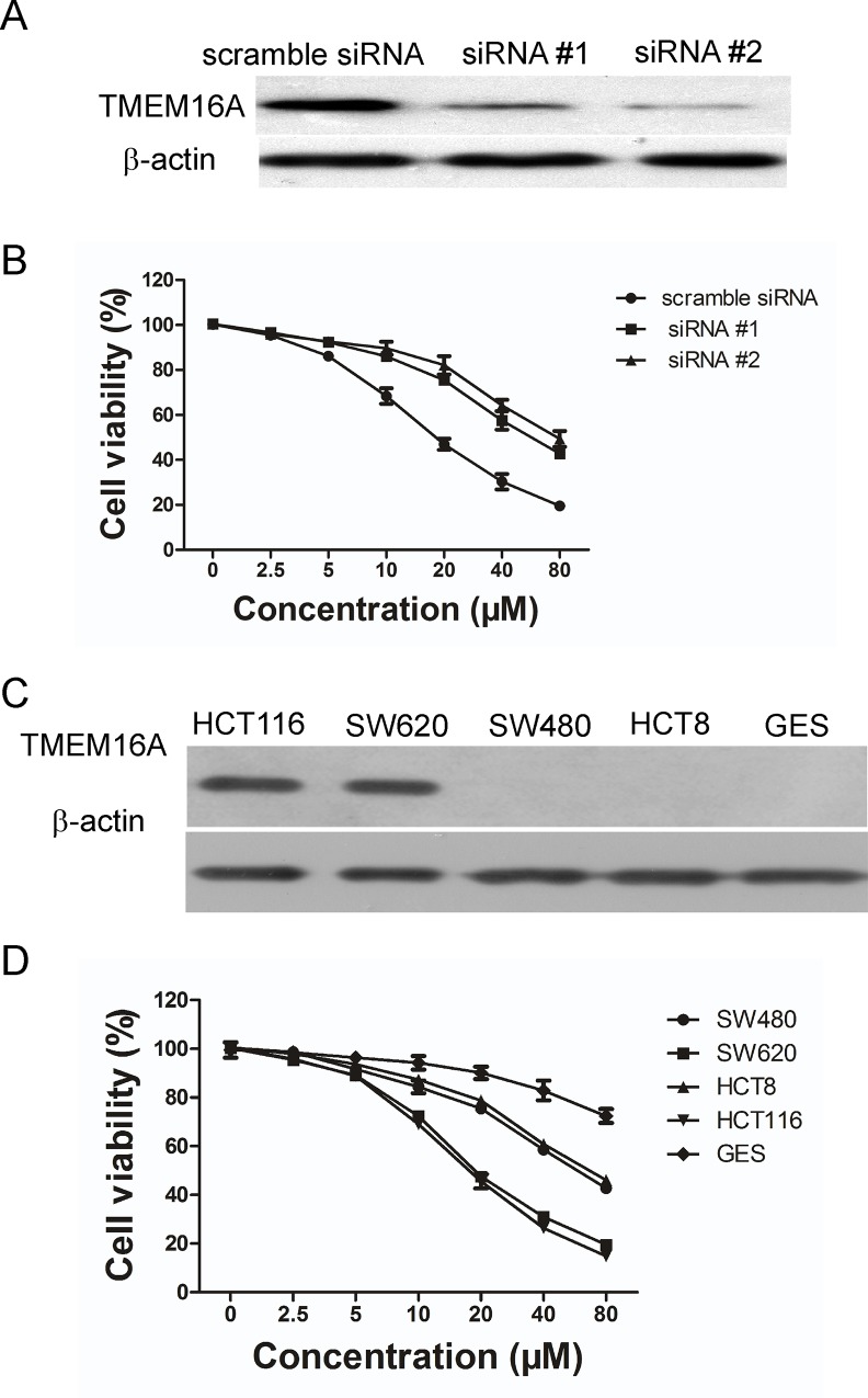
Fig 6. Involvement of TMEM16A in DP-induced growth inhibition.
A, Shown here is a western blot analysis of TMEM16A protein expression in SW620 cells that were transfected with siRNA #1 and #2. B, Effect of DP on SW620 cells after TMEM16A was knocked down, compared to scrambled siRNA. The viability of cells was determined by an MTT assay. Data are expressed as the mean ± SD (n = 5). DMSO served as the solvent control. C, TMEM16A expression was examined in the indicated cell lines by western blotting. Representative blots are shown. D, Effect of DP on growth of the indicated cell lines (mean ± SD, n = 4).

