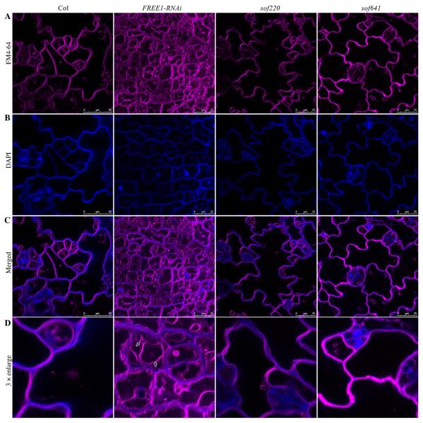
Fig. 3. Vacuole morphology analysis of representative FREE1-related sof mutants.
A: Vacuole morphology was analyzed using FM 4-64 dye staining. Compared to the wild type plants, fragmented vacuoles were observed in FREE1-RNAi plants. FREE1-related sof mutants sof220 and sof641 showed large central vacuoles. B: The short-time DAPI staining was used to visualize the cell shape. C: Merged result of FM 4-64 dye staining and DAPI staining. Scale bar, 25 μm. D: 3 × enlargement of merged images. Arrows indicated the fragmented vacuoles in FREE1-RNAi plants.

