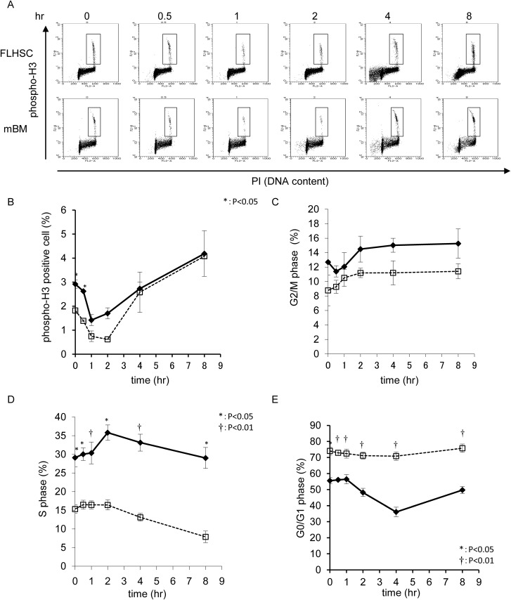
Fig 3.
(A) Cell-cycle distribution was monitored by a combination of phospho-histone H3 positivity and PI incorporation. Etoposide (10 mg/kg) was IP injected into E13.5 pregnant mice, and samples were analyzed at the indicated time points. A two-dimensional dot blot is shown. (B) Kinetics of phospho-histone H3 positivity indicating M phase percent, shown as a line graph. Bold line indicates FL-HSC, and broken line indicates mBM. (C) G2/M phase cell percent, (D) S phase cell percent, and (E) G0/G1 phase cell percent.

