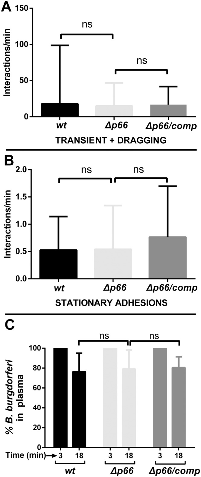
Fig 8. The effect of p66 deletion on vascular adhesion and clearance in a high- passage B. burgdorferi strain in BALB/c mice.

Non-infectious GFP-expressing B. burgdorferi wild type (GCB3212), Δp66 (GCB3214), and a strain where the wild-type p66 gene was reintroduced into the p66 deletion mutant (Δp66/comp, GCB3218) were injected into the jugular vein of BALB/c, 4x10⁸ spirochetes per mouse (n = 7/group). Over a period of up to 45 minutes, microvascular interaction rates A) (tethering + dragging), and stationary adhesions B) were enumerated in the knee joint-proximal tissue by intravital microscopy using spinning disk laser confocal microscopy as described in Materials and Methods. Blood vessels were stained with PE-conjugated PECAM-1 antibody. Statistical significance was analyzed using the non-parametric Kruskal-Wallis test; ns denotes not significant (P-values >0.05). C) Concentrations of B. burgdorferi in mouse plasma after iv inoculation. BALB/c mice were inoculated with B. burgdorferi through the tail vein as above and blood was withdrawn at 3 and 18 minutes post-inoculation (n = 7/group). Blood cells were allowed to settle overnight as described in Materials and Methods and spirochetes in the plasma were directly counted by dark-field microscopy. The change in spirochete concentration between 3 and 18 minutes was determined for each mouse as the percentage of spirochetes present at 18 minutes relative to the initial 3 minute time point. Statistical significance was analyzed using the non-parametric Kruskal-Wallis test; ns denotes not significant (P-values >0.05).
