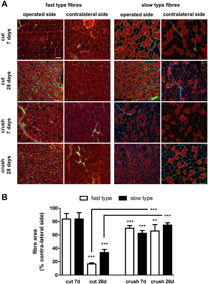
Fig 2. Fast and slow type gastrocnemius muscle fibre morphology.
(A) Transverse sections of contra-lateral and operated side muscles were stained with laminin antibody (green) and either fast type or slow type myosin heavy chain protein antibody (red). Samples shown are from animals undergoing sciatic nerve transection (cut) or crush injury 7 and 28 days after nerve injury. Scale bar = 50 μm. (B) Quantification of muscle fibre size. Computerised image analysis was used to calculate the mean ± SEM area (μm2) of fast type and slow type fibres in muscle obtained from the contra-lateral and operated sides of animals undergoing sciatic nerve transection (cut) and crush injury 7 and 28 days after insult. Data are expressed as percentage of the contra-lateral side. *P< 0.05, **P< 0.01, ***P< 0.001. Connecting bars also show ***P<0.001 for fast type and slow type fibres 28 days after cut and crush injury respectively.

