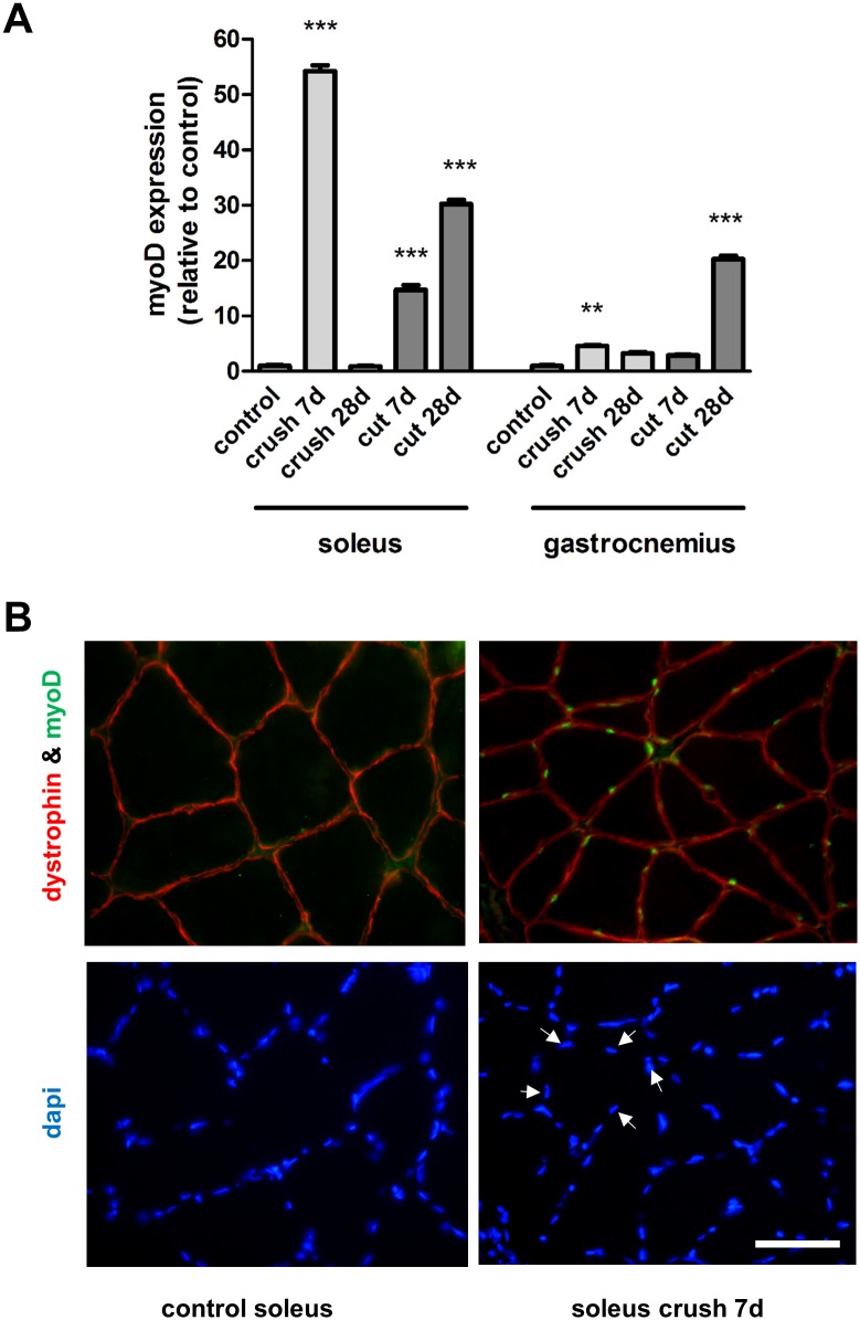
Fig 3. Expression of MyoD in muscles after nerve injury.
(A) The medial gastrocnemius muscles and the soleus muscles were harvested and fast frozen in liquid nitrogen for subsequent qRT-PCR analysis of myoD expression 7 and 28 days after nerve transection (cut) or crush injury. **P<0.01, ***P< 0.001 represents statistically significant difference to the respective control (unoperated group) muscles. Two-Way ANOVA indicates that the soleus and gastrocnemius muscles are significantly (P<0.001) different for each type of injury. (B) Control and soleus muscles harvested from animals 7 days after nerve crush injury were stained with antibodies directed against dystrophin (outline of muscle fibre) and myoD (green). Arrows highlight 5 representative nuclei (DAPI staining) which correspond with myoD positive staining.

