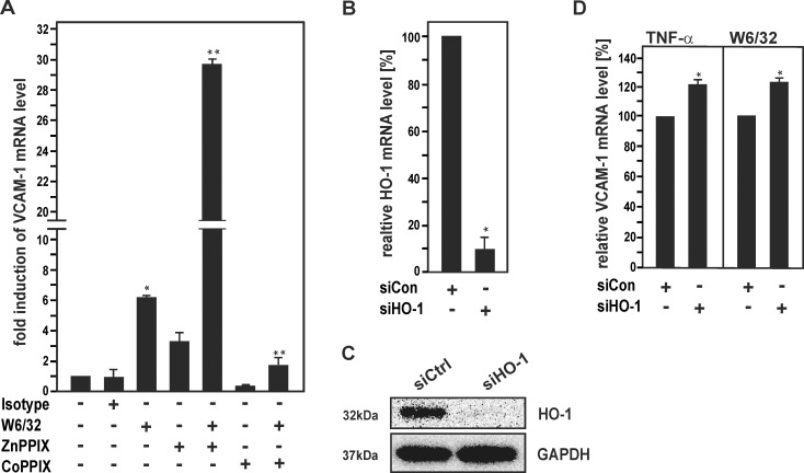
Fig 3. Pharmacological inhibition and siRNA-mediated knockdown of HO-1 reduce HLA I Ab-induced VCAM-1 expression in HUVECs.
(A) HUVECs were incubated with HLA I Ab W6/32 alone (10 μg/ml) and for 18 h in the presence of CoPPIX (5 μM) or ZnPPIX (5 μM), as indicated. Cells were lysed and subjected to RT-PCR analysis. The fold induction of VCAM-1 mRNA levels is shown. (B-D) HUVECs were transfected with siRNA for HO-1 or scrambled control siRNA. (B) mRNA expression was determined by RT-PCR analysis and relative levels of HO-1 mRNA are shown. (C) Protein expression was determined by Western blot analysis and probed sequentially with Abs against HO-1 and GAPDH. A representative of three independent experiments is shown. (D) Transfected HUVECs were treated with TNFα (15 ng/ml) or W6/32 for 18 h. Cells were lysed and subjected to RT-PCR analysis. The fold induction of mRNA levels is indicated. Bar graphs represent mean ± SEM from three independent experiments. * p≤ 0.05, significant differences treatment versus control; ** p≤ 0.05, W6/32 versus W6/32 plus CoPPIX/ ZnPPIX. Con, control.

