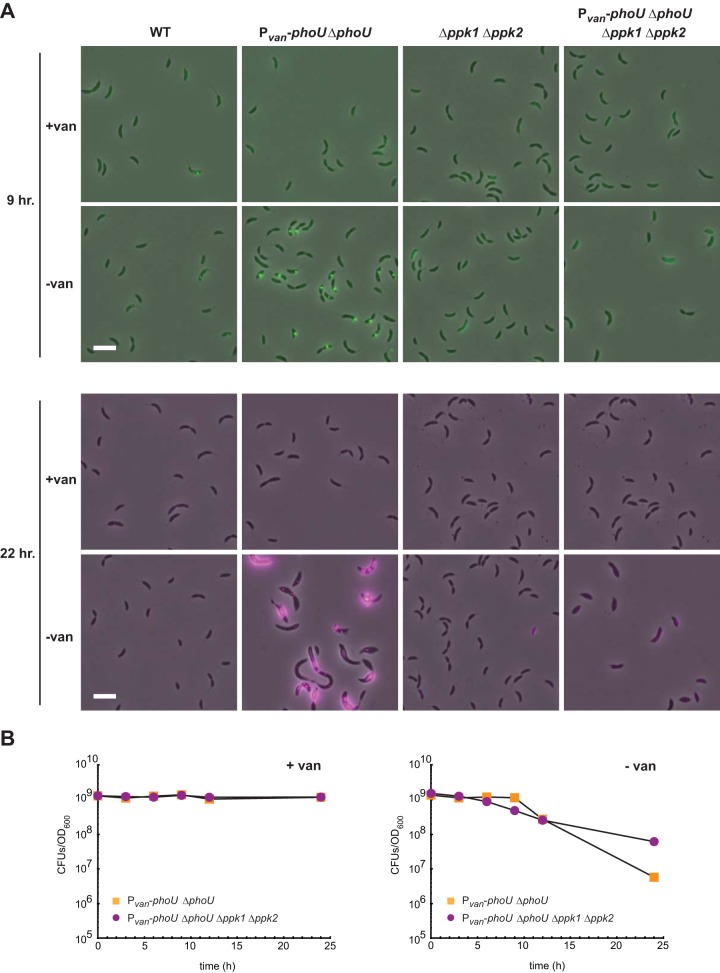
FIG 7.
Polyphosphate accumulates upon PhoU depletion in Caulobacter crescentus. (A) The strains indicated were grown either with or without vanillate for the times indicated. Cells were imaged by phase-contrast and epifluorescence microscopy using a filter set specific for DAPI-polyphosphate. Phase images were overlaid with the fluorescence image at 100% fluorescence excitation intensity (in green) for the 9-h time points and at a 30% excitation intensity (in magenta) for the 22-h time points. Bars, 2 μm. (B) Numbers of CFU for the strains indicated, with or without vanillate, are shown, with data points representing the averages of results from two replicates and given as the number of CFU in 1 ml normalized to an OD600 of 1.

