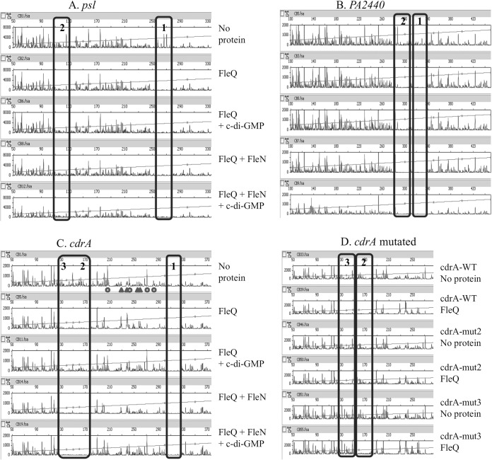
FIG 4.
FleQ binding does not require FleN or c-di-GMP. FAM-labeled DNA (0.45 pmol) was incubated with or without FleQ or FleN in the presence of ATP (10 μM) and in the presence or absence of c-di-GMP (500 μM) and then submitted to DNase I treatment (0.3 U) and analyzed by capillary electrophoresis. The fluorescence intensity (arbitrary units, ordinate) is plotted against the sequence length (bases, abscissa) of the fragment relative to the first base of the FAM-labeled primer. The horizontal line indicates the GS-500 internal size standard. The FleQ binding sites are boxed. (A) psl DNA (594 bp) was tested with 10 pmol of FleQ or FleN. (B) PA2440 DNA (522 bp) was tested with 50 pmol of FleQ or FleN. (C) cdrA fragment DNA (438 bp) was tested with 50 pmol of FleQ or FleN, and the locations of major putative DNA rearrangements are indicated by triangles (hypersensitive sites) or circles (hyposensitive sites). (D) The wild-type (WT) cdrA DNA fragment or cdrA fragments mutated in box 2 or 3 were tested with 50 pmol of FleQ.

