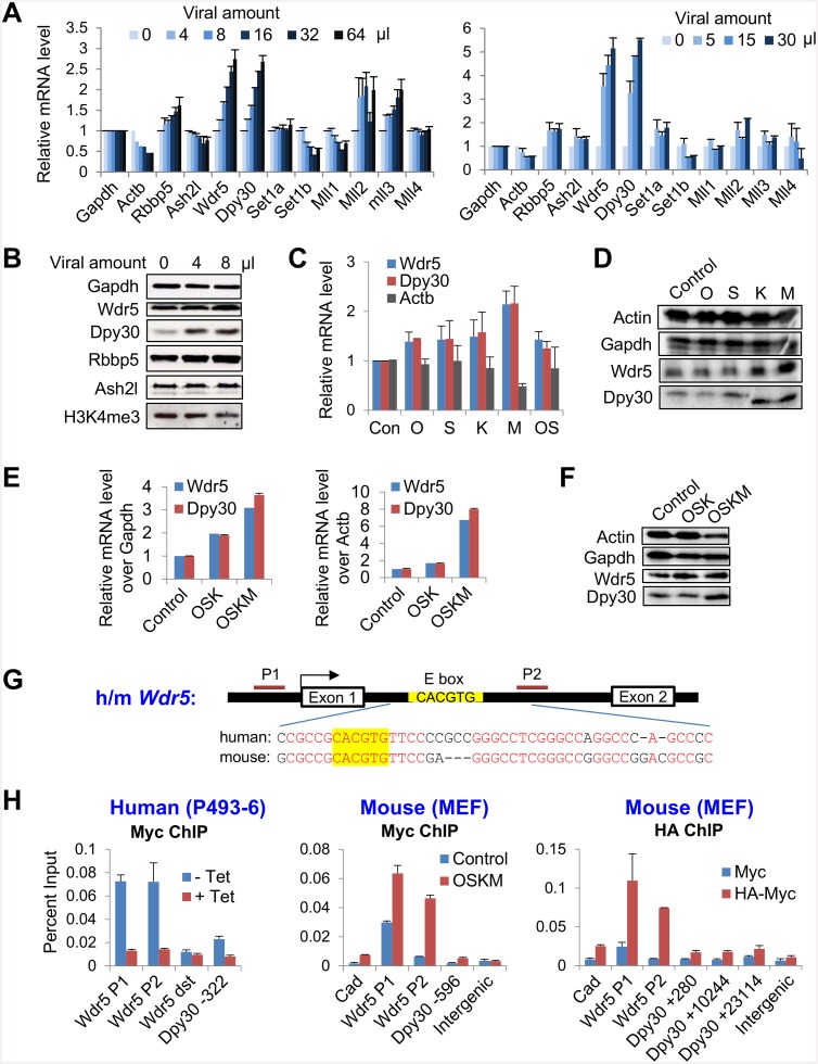
Fig 1. The reprogramming factors promote the expression of the core subunits of Set1/Mll complexes.
(A) MEFs were infected with increasing dose of FUW-M2rtTA and TetO-FUW-OSKM viruses in two independent infection experiments, and treated with doxycycline for 2 days. The mRNA levels of Set1/Mll complex subunits was determined by RT-qPCR and normalized by Gapdh. Average ± range of values from duplicate assays are plotted. (B) MEFs were infected with increasing dose of FUW-M2rtTA and TetO-FUW-OSKM viruses and treated with doxycycline for 2 days, and the expression of core subunits and global H3K4me3 were examined by western blotting. (C and D) MEFs were infected with virus expressing indicated individual reprogramming factor together with FUW-M2rtTA virus. MEFs infected with only FUW-M2rtTA virus was used as control (Con), and some MEFs were co-infected with TetO-FUW-Oct4 and TetO-FUW-Sox2 viruses (OS), in addition to FUW-M2rtTA. After induction with doxycycline for 2 days, the mRNA (C) and protein (D) levels of indicated Set1/Mll complex subunits were determined by RT-qPCR and normalized by Gapdh (C) and western blotting (D). Average ± SD from 3 independent infections are plotted in (C). (E and F) MEFs were infected with viral mixes that expressed individual Oct4, Sox2, Klf4 (OSK) or Oct4, Sox2, Klf4, and c-Myc (OSKM), in addition to FUW-M2rtTA, and MEFs infected with only FUW-M2rtTA virus was used as control. After induction with doxycycline for 2 days, the mRNA levels (E) of indicated core subunits were determined by RT-qPCR and normalized by Gapdh (E, left) or Actb (E, right). Average ± range of values from 2 independent infections are plotted. The indicated proteins were determined by western blotting (F). (G) Structure of human and mouse Wdr5 genes showing the highly conserved intronic sequences flanking the canonic E box. Identical residuals between human and mouse are in red. (H) Left: P493-6 cells were cultured in the absence (-Tet, Myc on) or presence of (+Tet, Myc off) tetracycline, and used for Myc ChIP followed by qPCR on indicated loci. Middle: MEFs were infected with only FUW-M2rtTA virus (control) or with FUW-M2rtTA and TetO-FUW-OSKM viruses (OSKM), induced with doxycycline, and used for Myc ChIP followed by qPCR on indicated loci. Right: MEFs were infected with FUW-M2rtTA and TetO-FUW-Myc virus (Myc) or FUW-M2rtTA and TetO-FUW-HA-Myc viruses (HA-Myc), induced with doxycycline, and used for HA ChIP followed by qPCR on indicated loci. Wdr5 dst, a Wdr5 downstream region. Cad is a previously established Myc target [63] and used as a positive control. Note that there are E boxes at +10088 and +23063 bp in the Dpy30 gene body. Average ± SD from triplicate assays are plotted, except for Myc ChIP in MEFs, for which Average ± range of values from duplicate assays are plotted. The differences between blue and red bars in all three panels are statistically significant (P<0.05 in 2-tailed Student’s t-test) for all loci except for the “Wdr5 dst” and the “Intergenic” sites.

