Figure 2.

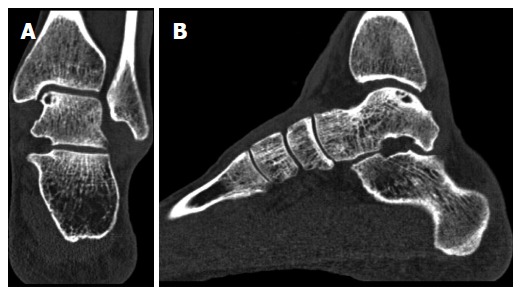
Coronal (A) and sagittal (B) computed tomography scans of a left ankle, showing an osteochondral defect of the posteromedial talar dome. Note the clear visualization of the cyst with an intact subchondral bone plate.

Coronal (A) and sagittal (B) computed tomography scans of a left ankle, showing an osteochondral defect of the posteromedial talar dome. Note the clear visualization of the cyst with an intact subchondral bone plate.