
Figure 5.
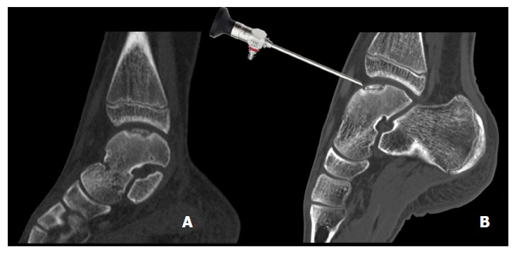
Sagittal computed tomography images of a 14-year-old patient with an osteochondral defect of the medial talar dome. Normal helical CT (A) and a CT made in full plantar flexion (B) showing arthroscopic accessibility. CT: Computed tomography.