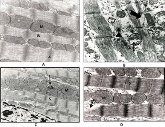
Figure 3.

Effect of flavored waterpipe exposed rat on heart ventricular tissue. A: ultrathin section of ventricular cardiomyocytes of air-exposed rat. M: mitochondrion with normal cristae, S: sarcomeres, Z: Z-line. Magnification:62500x. B: ultrathin section of ventricular cardiomyocytes of flavored waterpipe exposed rat. The thick arrows indicate a partial disruption of the myofibrils. Thin arrow indicates pleomorphic mitochondria with partially disrupted cristae. The triangle indicates deposited material. Magnification: 62500x. C: Ultrathin section of ventricular cardiomyocytes of flavored waterpipe exposed rat, showing enlarged mitochondria, irregularly arranged with partial disrupted cristae. Arrow indicates deposited material, M: mitochondrion, S: sarcomeres, N: nucleus. Magnification:25000x. D: ultrathin section of ventricular cardiomyocytes of flavored waterpipe exposed rat after the recovery period, showing partial recovery of cardiac muscle fiber. Enlarged mitochondria with partial disruption cristae. Arrow indicates a partial disruption of the myofibrils, M: mitochondrion, S: sarcomere. Magnification:62500x
