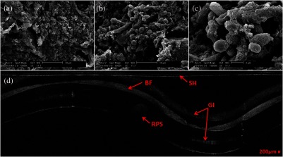
Fig. 3.
BF, biofilm; SH, probe sheath; RPS, radiopaque strip; and GI, ghost image; (a) SEM image of 120-h postintubation endotracheal tube showing the presence of extracellular polymeric substrate; (b) SEM image of same endotracheal tube in a different location; (c) SEM of same endotracheal tube focusing in on a cluster of cells; and (d) corresponding OCT image.

