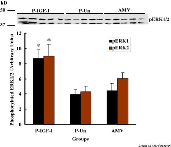
Figure 7.

Western blot analysis showing the level of phosphorylation of extracellular signal-regulated kinase-1/2 (pERK1/2) in mammary tissues from parous rats treated with insulin-like growth factor (IGF)-I (P-IGF-I), untreated parous rats (P-Un), and age-matched virgin rats (AMV). The IGF-I treatment (0.66 mg/kg body weight/day) was continued for 7 days before samples were collected. The protein samples (50 μg/lane) were electrophorized on 10% SDS-PAGE, transferred to PVDF membrane, and probed with antibody generated to phosphorylated form of human ERK1/2 (#9106; Cell Signaling Technology Inc., Beverly, MA, USA). Protein bands were detected using enhanced chemiluminescence reagents (upper image) and quantified using the ImageJ (version 1.24o; National Institutes of Health, Bethesda, MD, USA) image analysis program (bar chart). Values are expressed as mean ± standard error. *P < 0.05 versus P-Un and AMV.
