Figure 4.
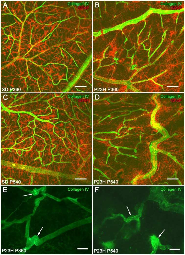
Superficial vascular plexus in SD and P23H rat retinas. Whole-mounted retinas from SD rats (A,C) and P23H rats (B,D–F) at P360 (A,B,E), and P540 (C,D,F), stained for collagen IV (green) and GFAP (red) in order to show the relationship between blood vessels and astrocytes, respectively, during retinal degeneration. During aging, blood vessels in P23H rat retinas (B,D) suffered alterations related to astrocyte changes that were unobserved in SD rats (A,C). Note the formation of blood vessel tangles at P360 (E, arrows) and P540 (F, arrows). Scale bar: 100 μm (A,D), 40 μm (E,F).

