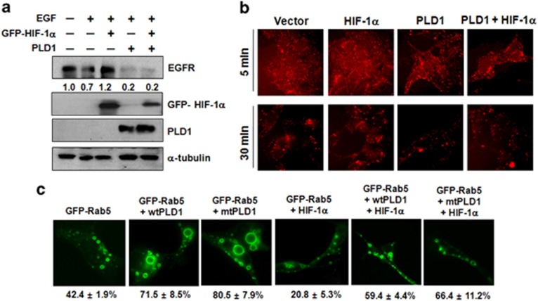
Figure 1.
PLD1 recovers HIF-1α-mediated inhibition of EGFR endocytosis via the Rab5/7 pathway. (a) HEK293 cells were transfected with GFP–HIF-1α and PLD1, then incubated in serum-free media for 14 h and stimulated with EGF (100 ng ml−1) for 1 h. The indicated proteins were analyzed by western blot. The values were normalized to that of α-tubulin and expressed relative to the control. (b) After HEK293 cells were transfected with GFP–HIF-1α and/or PLD1, they were treated with Alexa Fluor 555–EGF (20 ng ml−1) for the indicated times, fixed and examined by fluorescence microscopy. (c) HEK293 cells were transfected with GFP–Rab5 (Q79L), wtPLD1, mtPLD1 and HA–HIF-1α, then visualized by fluorescence microscopy. Data are representative of three independent experiments. EGFR, epidermal growth factor receptor; HA, hemagglutanin; HIF-1α, hypoxia-inducible factor-1α PLD, phospholipase D.

