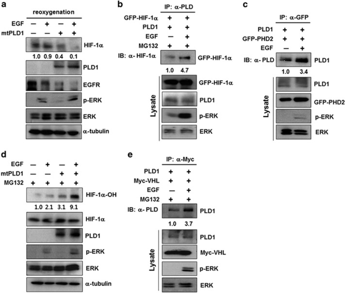
Figure 3.
PLD1 promotes EGFR endocytosis by destabilizing HIF-1α protein via the PHD2/VHL-mediated pathway. (a) HEK293 cells were transfected with mtPLD1, then incubated under hypoxia. After hypoxia for 4 h, the cells were reoxygenized and treated with EGF (100 ng ml−1) for 30 min. The lysates were then analyzed by immunoblot. The values were normalized to that of α-tubulin and expressed as a fold of the control. (b and c) HEK293 cells cotransfected with the indicated constructs and stimulated with EGF (100 ng ml−1) for 30 min in the presence MG132. The lysates were immunoprecipitated with anti-PLD (b) or anti-GFP (c) and immunoblotted with the indicated antibodies. The values were normalized against that of α-tubulin and expressed as a fold of the control. (d) Immunoblot assay of the indicated proteins in HEK293 cells transfected with mt PLD1 and stimulated with EGF in the presence MG132. The hydroxylated values were normalized to total HIF-1α and expressed as a fold of the control. (e) HEK293 cells transfected with mt PLD1 and stimulated with EGF in the presence MG132. The lysates were immunoprecipitated and immunoblotted with the indicated antibodies. Data are representative of three independent experiments. EGFR, epidermal growth factor receptor; HIF-1α, hypoxia-inducible factor-1α GFP, green fluorescent protein; PLD, phospholipase D; VHL, von Hippel–Lindau.

