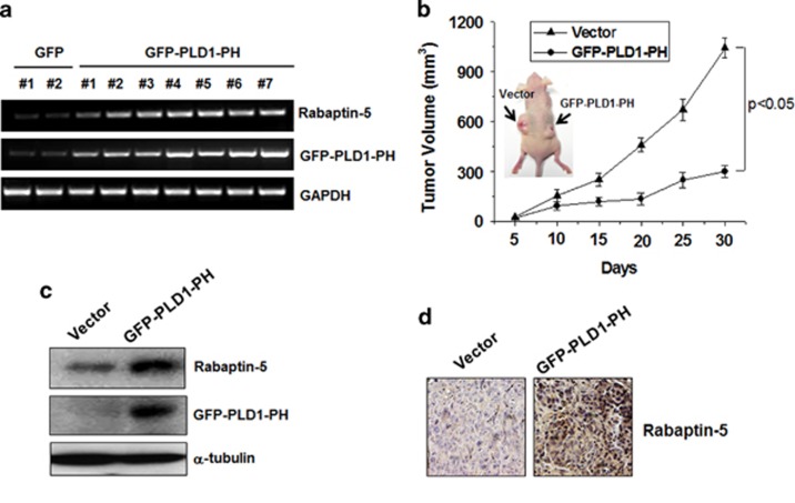
Figure 5.
PLD1-PH attenuates tumor progression. (a) RT–PCR assay of rabaptin-5 mRNA in HT29 cells stably transfected with PLD1-PH. (b) HT29 cells expressing empty vector or PLD1-PH cells were xenografted on the left and right flanks of nude mice, respectively (arrows), then analyzed for tumor volume. Photographs show the representative tumors. Data were expressed as the mean±s.d. of seven different mice. (c) Immunoblot analysis of rabaptin-5 in tumor tissues from the xenografted mice injected with vector and PLD1-PH-expressed HT29 cells. Data are representative of three independent experiments. (d) Immunohistochemical staining of rabaptin-5 in the tumor tissues of xenografted mice injected with vector and PLD1-PH-expressed HT29 cells. Data are representative of three independent experiments. PH, pleckstrin homology; PLD, phospholipase D; RT–PCR, reverse transcription–PCR.

