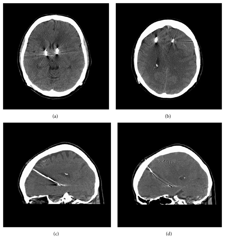
Figure 1.
(a) CT head demonstrates left to right brain shift. (b) CT head demonstrates isodense pathology along left convexity. (c) CT head sagittal view demonstrates R DBS lead with relative normal position. (d) CT head sagittal view demonstrates L DBS lead that has bowed compared to the R DBS lead.

