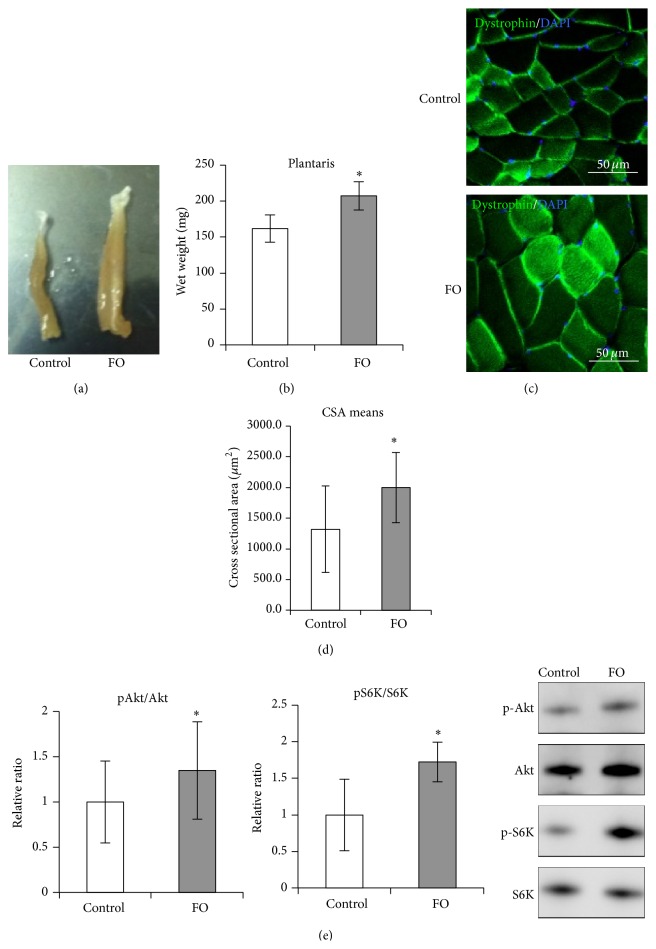
Figure 1.
Skeletal muscle hypertrophy following functional overload. (a, b) Change in muscle weight following functional overload. A photograph of plantaris muscles isolated from control and FO groups (a) and a graph representing wet weight of plantaris in each group (b). (c, d) Change in cross-sectional area of plantaris following functional overload. Representative merged images of immunohistochemistry staining for Dystrophin (green) with DAPI from control and FO groups (c) and a graph plotting the means of cross-sectional area in each group (d) are shown. (e) Representations of Akt (Ser473) phosphorylation levels (left) and p70S6K (Thr389) phosphorylation levels (center), as detected by Western blot analysis. The typical blot patterns are described in the right panels. Phosphorylation levels were calculated to divide the signal of the phosphorylated form against the total protein expression for Akt or S6K. The relative ratio, normalized to the signal observed for the control group, is shown. All values are expressed as the mean ± SDM (n = 5). Significant differences: ∗compared to control group (P < 0.05).

