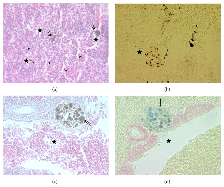
Figure 6.
(a) Splenic tissue of Dicentrarchus labrax L. organized in areas of red pulp (r) and white pulp (w); the last consisting of ellipsoids, MØ (arrow), and MMC (double arrow) (H&E, 100x). ((b), (c), (d)) At higher magnification (400x) MØ and MMC appear distributed above all near vessels (star) (Anae, Mallory, and Perls-Van Gieson, resp.).

