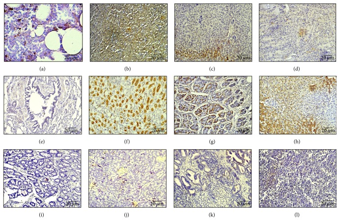
Figure 3.
The distribution of MSCs with intra-arterial infusion in femoral head and vital organs. Immunohistochemical staining of BrdU-positive MSCs in different tissues at eight weeks after transplantation was observed under a light microscope. (a) Femoral head. (b) Heart; (c) liver; (d) spleen; (e) lung; (f) kidney; (g) stomach; (h) gallbladder; (i) small bowel; (j) pancreas; (k) prostate; (l) testicle. Scale bar 20 μm. Particularly, BrdU strongly positive MSCs were significantly located in the tissues of the kidney, gallbladder, and liver.

