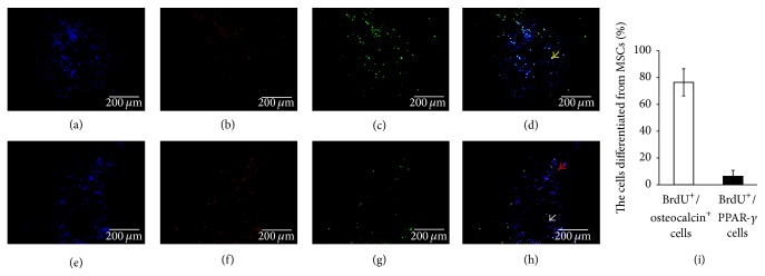
Figure 4.
Osteogenic and adipogenic differentiation of MSCs with intra-arterial infusion in necrotic field of femoral head. BrdU (green) and osteocalcin (red) were analyzed by double-immunofluorescent analysis in the necrotic region of MSC-transplanted femoral heads. (a) DAPI (blue); (b) BrdU (red); (c) osteocalcin (green); (d) an overlay of (a), (b), and (c); (e) DAPI (blue); (f) BrdU (red); (g) PPAR-γ (green); (h) an overlay of (e), (f), and (g). Most of the BrdU-positive MSCs in the necrotic region of MSC-transplanted femoral heads costained together with osteocalcin but a few cells costained together with PPAR-γ. Yellow arrow shows osteoblasts differentiated from MSCs, red arrow shows adipocytes differentiated from MSCs, and grey arrow shows BrdU-positive cells which did not differentiate to lipocytes. Scale bar 200 μm. BrdU+/osteocalcin+ cells in MSCs group (76.42 ± 10.14%) were significantly higher than BrdU+/PPAR-γ + cells over total BrdU+ cells (6.36 ± 4.41%) after eight-week treatment (p < 0.05) (i).

