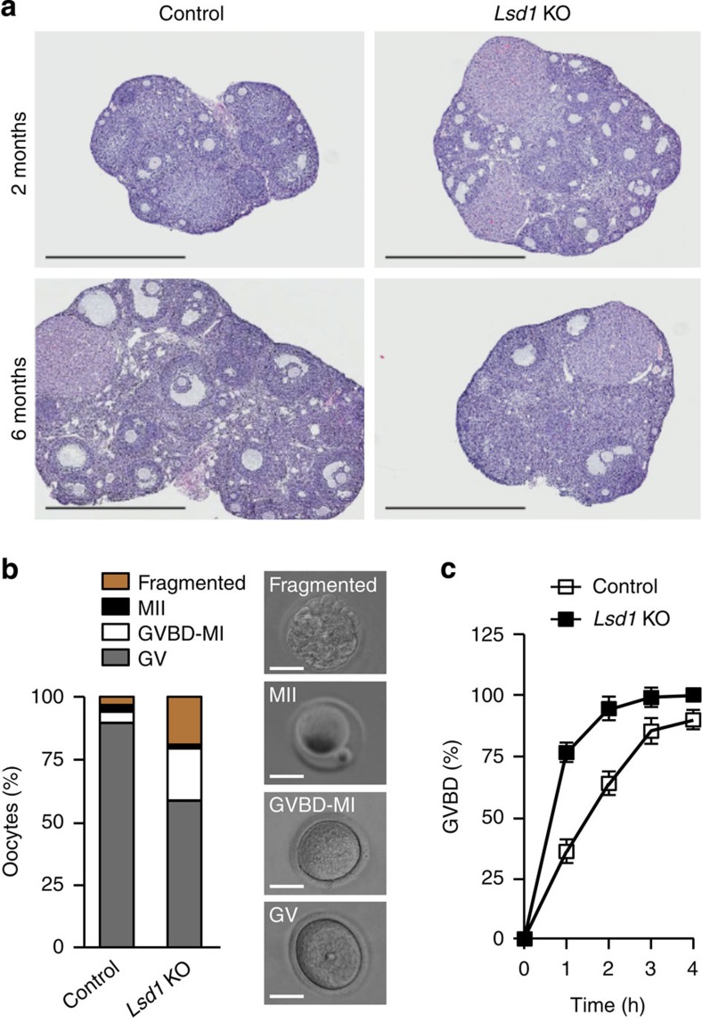
Figure 2. LSD1 depletion in oocytes results in precocious meiotic resumption.
(a) Haematoxylin and eosin (H&E) staining of ovarian sections. Shown are representative images of ovaries from 2-month- and 6-month-old control and Lsd1 KO mice. Scale bars, 1 mm. (b) Fully grown GV oocytes isolated from ovaries were cultured in the presence of 200 μM IBMX for 20 h and classified as being GV arrested (based on the presence of germinal vesicle; grey bar), GVBD-MI (based on the absence of both germinal vesicle and polar body; white bar), MII (based on the presence of a polar body; black bar) or fragmented (yellow bar). The average proportion of oocytes at each stage from three experiments is plotted as a percentage of the total (left). Examples of GV, GVBD-MI, MII and fragmented oocytes are shown (right). Scale bars, 50 μm. (c) Fully grown GV oocytes were collected in M2 medium containing IBMX and, following IBMX washout, the oocytes were cultured in the absence of IBMX and examined hourly to determine GVBD rates. The data at each time point represent the mean±s.e.m. of three experiments.

