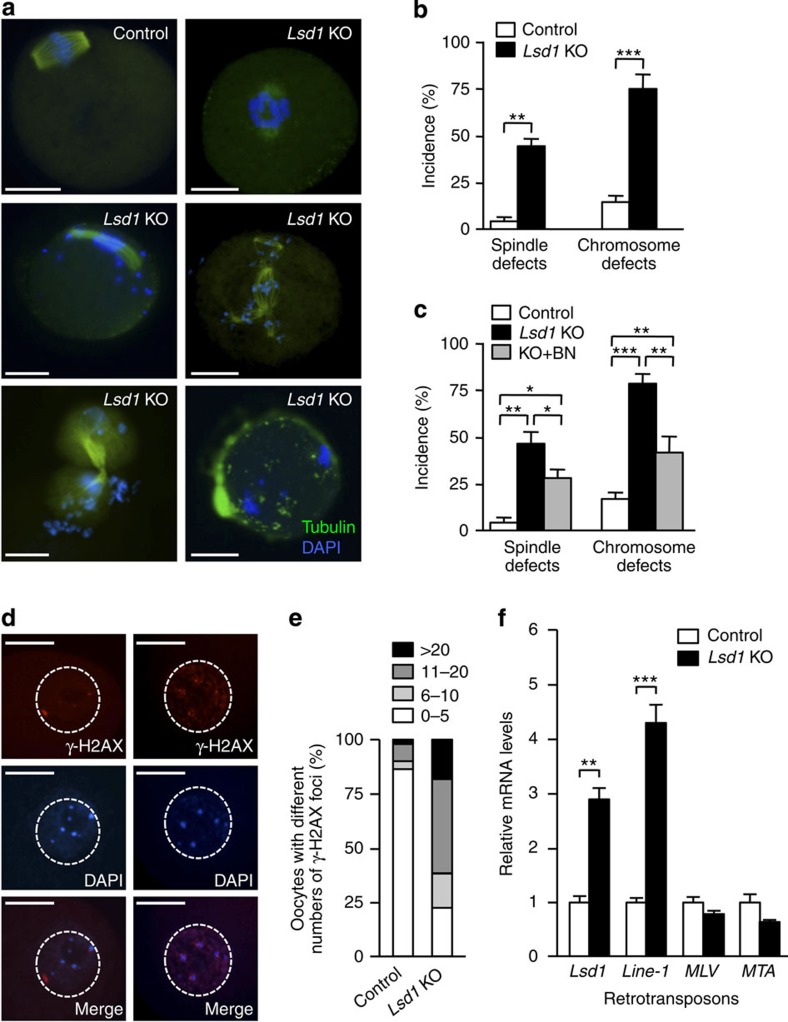
Figure 6. Lsd1 KO oocytes exhibit chromosomal defects and DNA damage.
(a,b) GV oocytes were collected from the ovaries of PMSG-primed females, cultured in maturation medium for 6 h, and then immunostained for α-tubulin (green) and DNA (blue) to examine spindle morphology and chromosome alignment. (a) Representative IF images showing normal spindle and chromosome structures in control oocytes and common abnormalities in Lsd1 KO oocytes. Scale bars, 25 μm. (b) Frequencies of spindle and chromosomal abnormalities in control and Lsd1 KO oocytes (mean±s.e.m. from three experiments). Statistical comparisons of values were made using multiple t-test. **P<0.01; ***P<0.001. (c) Effect of CDC25 inhibition on spindle and chromosome phenotypes in Lsd1 KO oocytes. GV oocytes were collected and cultured in maturation medium for 1 h to induce GVBD and then further cultured either with or without the CDC25 phosphatase inhibitor BN82002 (BN) for 6 h. Shown are frequencies of abnormal spindle and chromosome defects in oocytes of the indicated groups (mean±s.e.m. from three experiments). Statistical comparisons of values were made using one-way ANOVA. *P<0.05; **P<0.01; ***P<0.001. (d,e) GV oocytes were examined for DNA double-strand breaks (DSBs) with anti-γ-H2AX staining. (d) Representative γ-H2AX (red), DAPI (blue) and merged images of control (left) and Lsd1 KO (right) oocytes. The nuclei are circled. Scale bars, 25 μm. (e) The proportions of oocytes with various numbers of γ-H2AX foci in the nuclei are shown. (f) Quantitative RT–PCR analysis of retrotransposon transcripts in control and Lsd1 KO oocytes. IAP, intracisternal A particles; Line-1, long interspersed nuclear element-1; MLV, murine leukaemia virus; MTA, mouse transposon A. Statistical comparisons of values were made using multiple t-test. **P<0.01; ***P<0.001.

