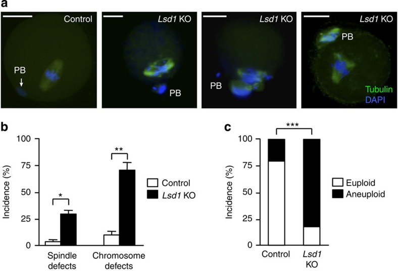
Figure 7. Lsd1-null MII oocytes exhibit chromosomal defects and aneuploidy.
(a,b) Oocytes were collected from the oviducts 16 h post-hCG injection and then immunostained for α-tubulin (green) and DAPI (blue) to examine spindle morphology and chromosome alignment. (a) Representative IF images showing normal spindle and chromosome structures in control MII oocytes and common abnormalities in Lsd1 KO MII oocytes. PB, polar body. Scale bars, 25 μm. (b) Frequencies of abnormal spindle and chromosome defects in MII oocytes of the indicated groups (mean±s.e.m. from three experiments). Statistical comparisons of values were made using multiple t-test. *P<0.05; **P<0.01. (c) Chromosome spreads of MII oocytes from control and Lsd1 KO mice were performed, and chromosome numbers counted. Shown are the percentages of euploid (20 chromosomes) and aneuploid (fewer or more than 20 chromosomes) MII oocytes from control and Lsd1 KO mice. Statistical comparisons of values were made using unpaired t-test. ***P<0.001.

