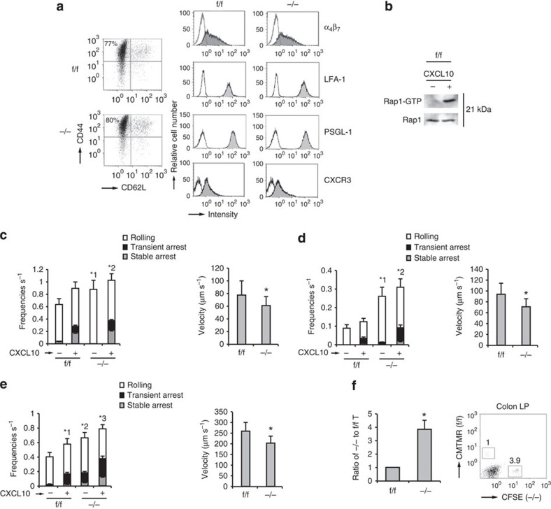
Figure 4. Rap1 deficiency enhances rolling of TEM cells.
(a) Representative expression profiles of CD44 and CD62L, α4β7, LFA-1, PSGL-1 and CXCR3 in CD4+T cells from f/f or −/− mice, cultured under TH17-polarizing conditions. (b) f/f TEM cells were incubated at 37 °C at 15 s in the presence or absence of CXCL10. Cell lysates were pulled down with GST-RalGDS-RBD and immunoblotted with anti-Rap1. (c; Left) f/f or −/− TEM cells were perfused on LS12 cells expressing P-selectin and mouse ICAM-1 ±CXCL10. Adhesion of more than 100 cells was measured. *1P<0.002, *2P<0.05 versus total frequency of the corresponding f/f TEM cells. (Right) The rolling velocity of f/f or −/− TEM cells without CXCL10. *P<0.03 versus f/f TEM cells. (d; Left) f/f or −/− TEM cells were perfused on LS12 cells expressing mouse MadCAM-1 ±CXCL10. Adhesion of more than 100 cells was measured. *1P<0.001, *2P<0.002 versus total frequency of the corresponding f/f TEM cells. (Right) The rolling velocity of f/f or −/− TEM cells without CXCL10. *P<0.01 versus f/f TEM cells. (e; Left) f/f or −/− TEM cells were perfused on plates immobilized with MadCAM-1 ±CXCL10. Adhesion of more than 100 cells was measured. *1P<0.02 versus unstimulated f/f TEM cells. *2P<0.01, *3P<0.03 versus total frequency of the corresponding f/f TEM cells. (Right) The rolling velocity of f/f or −/− TEM cells on MadCAM-1 without CXCL10. *P<0.005 versus f/f TEM cells. (f) Homing assay of TEM cells into the colon. (Left) f/f or −/− TEM cells labelled with CMTMR or CFSE were injected into normal mice. After 10 h, cells from the colon LP of recipient mice were analysed using flow cytometry. The ratios of −/− to f/f TEM cells are shown (n=3 experiments). (Right) Flow-cytometry profile of colon LP in the recipient mice. Numbers indicate the ratio of −/− to f/f TEM cells. All data show the means±s.e.m. or are representative of three independent experiments.

