
Figure 2. The surface charge distribution of the peptide-receptor complex structure based on Poisson-Boltzmann equation.
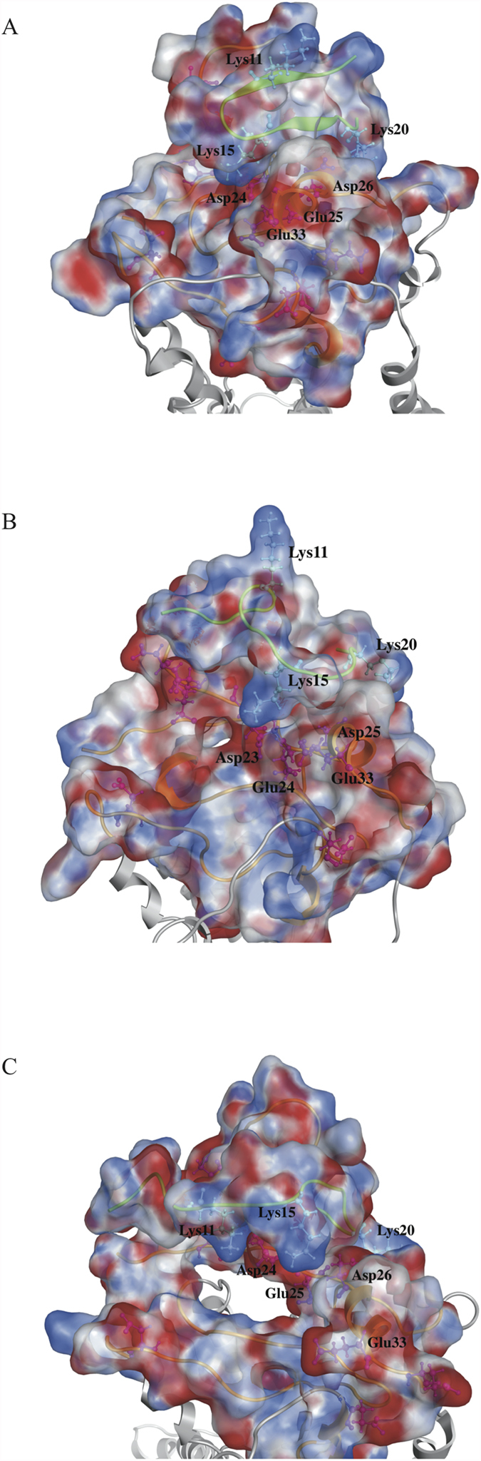
The three peptides ((A) p_wt14 (B) p_wt16 (C) p_wt18) still bind to the groove of N-terminal domain of CXCR1 during the 50 ns MD simulations with positively charged lysine residues facing the negatively charged groove region of CXCR1 indicating that electrostatic interactions dominate the initial binding. Side chains of positively charged residues are represented as light blue color while that of negatively charged residues are represented as pink color.