
Figure 3. The MM/PBSA binding free energy calculations for various peptides of CXCL8 binding to CXCR1.
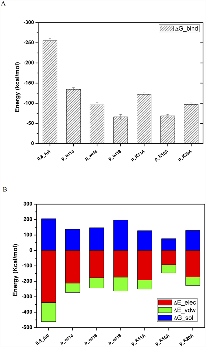
(A) For wild peptides with different lengths (p_wt14, p_wt16 and p_wt18) and mutant peptides (p_K11A, p_K15A and p_K20A). (B) The detailed analysis of the components of binding free energies shows that electrostatic interactions dominate the binding (red color), followed by solvation free energies (blue color) and van der Waals (VDW) interactions (green color).