
Figure 7. Comparison of various mutant peptides to inhibit CXCL8 induced monocyte adhesion to HMEC-1.
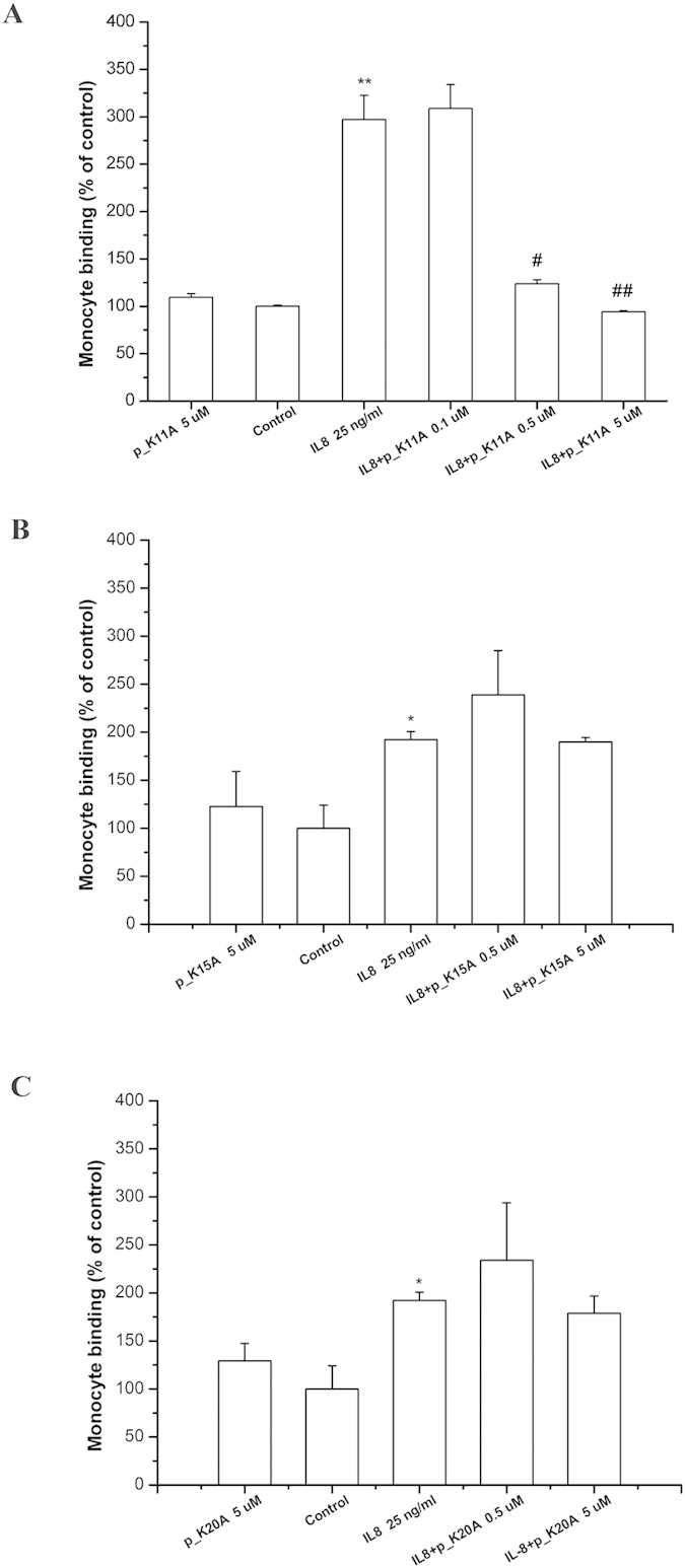
(A) Mutant peptide p_K11A (B) Mutant peptide p_K15A (C) Mutant peptide p_K20A. HMEC-1 was pretreated with various concentrations of the mutant peptide for one hour, and then stimulated with 25 ng/ml CXCL8 for 18 hours. Adhesion of fluorescent THP-1 cells was photographed by fluorescent microscopy and calculated. “Control” means that only the culture medium (without peptides) is incubated with cells. Values are mean ± SD from three independent experiments. (**P < 0.01) as compared to control; (#P < 0.05) and (#P < 0.01) as compared to cells stimulated with CXCL8 in the absence of peptides; (*P < 0.05) as compared to control.