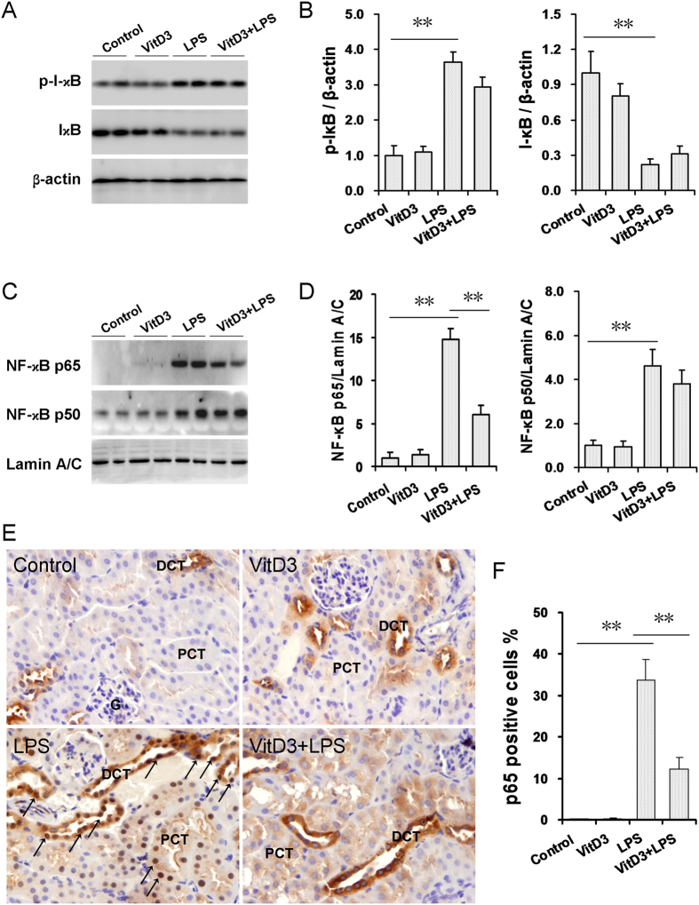
Figure 5. Effects of VitD3 pretreatment on LPS-activated renal NF-kB signaling. In LPS group, mice were i.p. injected with LPS (1.0 mg/kg).
In VitD3 + LPS group, mice were pretreated with three doses of VitD3 (25 μg/kg) at 48, 24 and 1 h before LPS injection. Kidneys were collected 1 h after LPS injection. (A,B) Renal I-κB and p-IκB were measured using immunoblots. All experiments were duplicated for four times. (A) A representative gel for p-IκB (upper panel), I-κB (middle) and β-actin (lower panel) was shown. (B) p-IκB/β-actin and I-κB/β-actin. All data were expressed as means ± S.E.M. (n = 8). **P < 0.01. (C,D) Renal nuclear NF-κB p65 and p50 subunits were measured using immunoblots. All experiments were duplicated for four times. (C) A representative gel for NF-κB p65 (upper panel), p50 (middle) and lamin A/C (lower panel) was shown. (D) p65/ lamin A/C and p50/ lamin A/C. All data were expressed as means ± S.E.M. (n = 8). **P < 0.01. (E) Renal nuclear NF-kB p65 translocation was analyzed using IHC. Representative photomicrographs of renal histological specimens from mice treated with NS, VitD3, LPS (1H) and VitD3 + LPS are shown. Original magnification × 400. Nuclear NF-kB p65 translocation was mainly observed in the nuclei of the distal convoluted tubules (arrows). DCT: distal convoluted tubule; PCT: proximal convoluted tubule; G: glomerulus. (F) Percent of p65 positive cells among different groups. All data were expressed as means ± S.E.M (n = 10). **P < 0.01.

