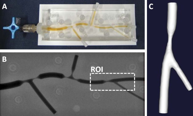
Fig 1. Overview of the 3D reconstruction steps of a phantom model with bi-plane angiography.
A: Original phantom model filled with contrast agent. B: An angiography recording of the phantom. A region of interest (ROI) is indicated around a section that can be regarded as mildly stenosed. C: Final 3D reconstruction.

