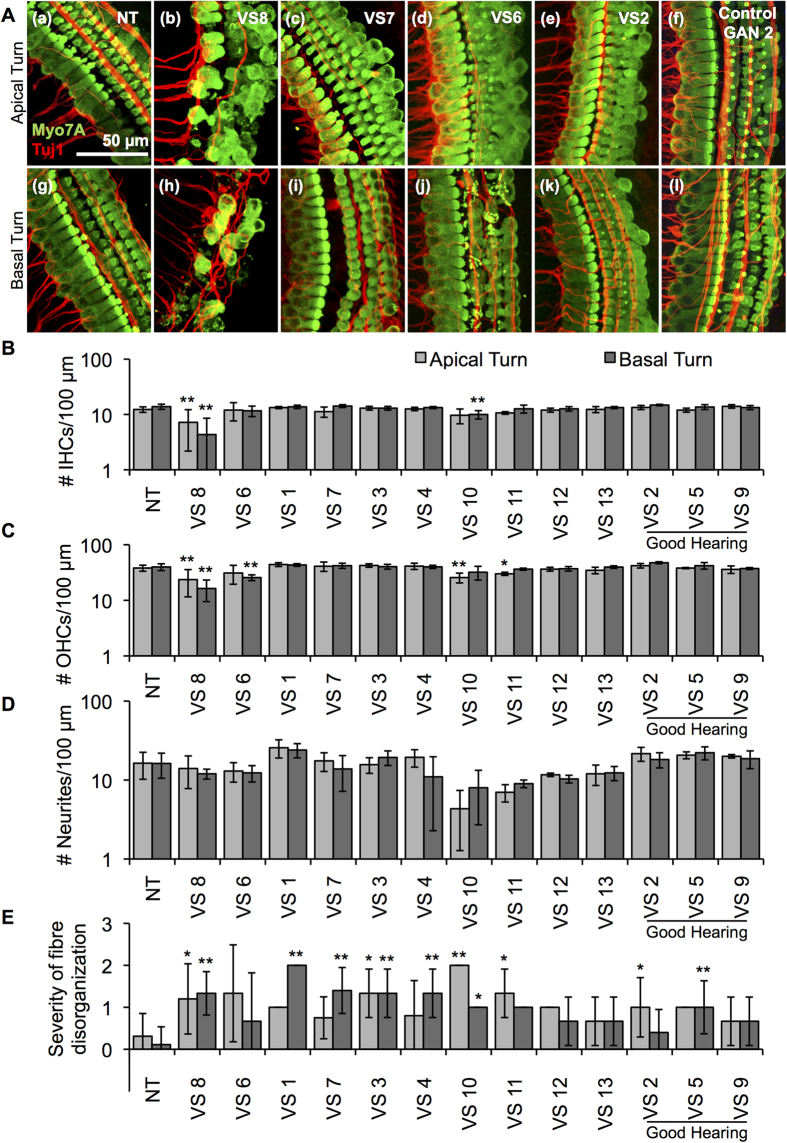
Figure 2. Application of human VS secretions onto murine cochlear explant cultures leads to hair cell and neurite loss.
(A). Representative images for cochlear explants receiving (a) no treatment (NT, n = 28 different explants), incubated with (b) VS8 (n = 5 different explants), (c) VS7 (n = 4 different explants), (d) VS6 (n = 3 different explants), (e) VS2 (n = 5 different explants) and (f) control GAN2 (n = 3 different explants) secretions are shown for the apical turns, and (g) NT (n = 26 different explants), (h) VS8 (n = 6 different explants), (i) VS7 (n = 5 different explants), (j) VS6 (n = 3 different explants), (k) VS2 (n = 5 different explants) and (l) control GAN2 (n = 4 different explants) secretions for the basal turn. Myo7A (green) marks hair cells and Tuj1 (red) marks neurites. Scale Bar = 50 μm applies to all images. (B). Number of inner hair cells (IHCs), (C). outer hair cells (OHCs), (D). neurites, and (E). severity of fibre disorganization are shown for a 100 μm length within the apex (light grey columns) and basal turn (dark grey columns) cochlear explants treated with NT and secretions from 13 different tumours. *p < 0.05, **p < 0.01. Quantified data after treatment with control GAN secretions are shown in Supplementary Figure 1.

