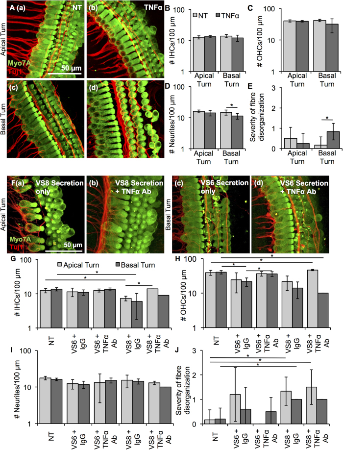
Figure 4. TNFα application onto cochlear explants induced mild damage.
(A). Representative images for cochlear explants receiving no treatment (NT, a, c, n = 6 different explants) or incubated with TNFα (b, d, n = 4–6 different explants) are shown for the apical (a, b) and basal (c, d) turn. Myo7A (green) marks hair cells and Tuj1 (red) marks neurites. Scale Bar = 50 μm applies to all images. (B). Number of inner hair cells (IHCs), (C). number of outer hair cells (OHCs), (D). number of neurites, (E). severity of fibre disorganization are shown for a 100 μm length within the apical and basal turn explants for NT (light grey columns) and TNFα-treated (dark grey columns). *p < 0.05. (F). Representative images for cochlear explants receiving VS secretions only (a, c, n = 2 different tumours) or incubated with VS secretions with TNFα antibody (b, d, n = 2 different tumours) are shown for the apical (a, b) and basal (c, d) turn. Myo7A (green) marks hair cells and Tuj1 (red) marks neurites. Scale Bar = 50 μm applies to all images. (G). Number of inner hair cells (IHCs), (H). number of outer hair cells (OHCs), (I). number of neurites, (J). severity of fibre disorganization are shown for a 100 μm length within the apical and basal turn explants treated with VS secretions only (light grey columns) and VS secretions with TNFα antibody (dark grey columns). *p < 0.05.

