
Figure 6.
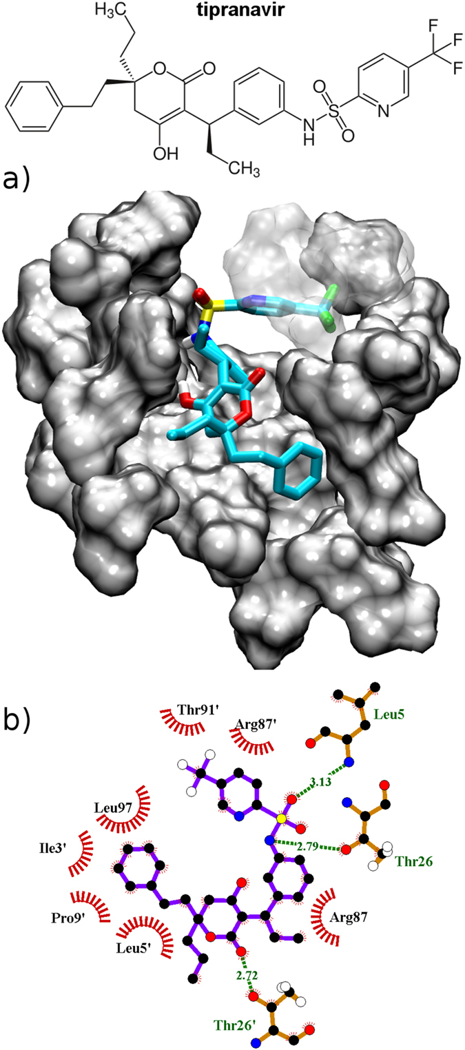
(a) Binding of tipranavir to the new pocket in conformation S2C. For the sake of clarity some protease residues are shown as transparent. The color code is as follows: C atoms - cyan, N atoms - blue, O atoms - red, F atoms - green and S atoms - yellow. (b) Tipranavir interactions with the binding site represented employing the software Ligplot+60.