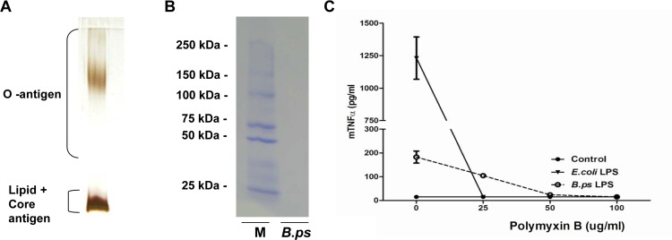
Fig 1. Successful extraction and purification of B.pseudomallei-LPS.
LPS from B.pseudomallei 1026b was purified by a modified hot phenol-water extraction method including proteinase K treatment. 10 μl of LPS (0.5 mg/ml) was fractionated by SDS-PAGE electrophoresis followed by silver staining (A) which showed the characteristic ladder pattern of LPS banding of Gram-negative bacteria, without any indications of protein contamination (detection limit of 0.5–5 ng) and Coomassie blue staining (B) which demonstrated no protein contamination as well (detection limit 50 ng). Contamination of the extracted LPS was also assessed by the addition of LPS-binding Polymyxin B (PMB) (C). For this purpose, the murine alveolar macrophage cell line MH-S was stimulated with RPMI 1640 medium [56], LPS of E. coli 0111:B4 or B.pseudomallei 1026b and incubated with increasing concentrations of PMB for 6h, followed by TNF-α measurement in the supernatant. Results are representative for three independent experiments. (M = ladder, B.ps = B.pseudomallei-LPS)

