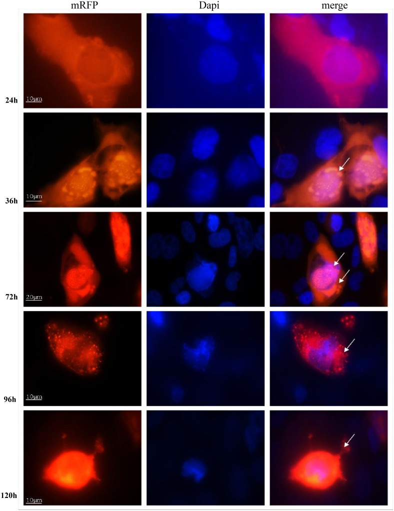
Fig 7. Capsid pIX intracellular expression.
U251 cells were infected at an MOI of 1 VP/cell and then fixed at the times indicated with PBS containing 10% formalin, and mounted with Vectashield with DAPI. Fluorescent signal for pIX-RFP was detected by fluorescent microscopy (all magnifications at 1,000 × except 72h at 600 ×). Arrows indicate virus particle specs in the cytoplasm by 24 hpi, in the nucleus and vacuoles by 36 hpi; viral specs released from the nucleus into the cytoplasm by 96 hpi, and released from the cell by 120 hpi.

