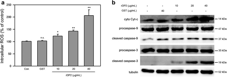
Fig. 2.

rDP2 increased intracellular ROS and cytosolic cytochrome c and induced activation of caspase-9 and caspase-3 in BEAS-2B cell. Cells were treated with rDP2 at indicated concentrations for 24 h, and then the cells were subjected to a intracellular ROS assay or b immunoblotting for indicated proteins. Three independent experiments were performed for statistical analysis. *p < 0.05 and **p < 0.01 as compared to GST treatment (40 μg/mL, 24 h). Signals of tubulin were used as internal control
