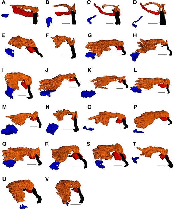
Fig. 5.

3D-reconstructions of the head glands of female Philanthinae. The postpharyngeal gland (PPG) is shown in orange and red; the mandibular gland (MG) is shown in blue. Note that for each species only the right part of the head capsule is shown. a Aphilanthops frigidus; b Clypeadon laticinctus; c Cerceris arenaria; d Cerceris quinquefasciata; e Philanthinus quattuordecimpunctatus; f Trachypus flavidus (note that for this species the MG could not be reconstructed based on the available serial histological sections); g Trachypus elongatus; h Trachypus boharti; i Philanthus venustus; j Philanthus triangulum diadema; k Philanthus capensis; l Philanthus loefflingi; m Philanthus rugosus; n Philanthus melanderi; o Philanthus coronatus; p Philanthus bicinctus; q Philanthus ventilabris; r Philanthus multimaculatus; s Philanthus barbiger; t Philanthus gibbosus; u Philanthus albopilosus; v Philanthus psyche. Color code: orange, upper part of the PPG; red, lower part of the PPG; blue, MG; black, pharynx. Scale bars = 0.25 mm
