Figure 2.

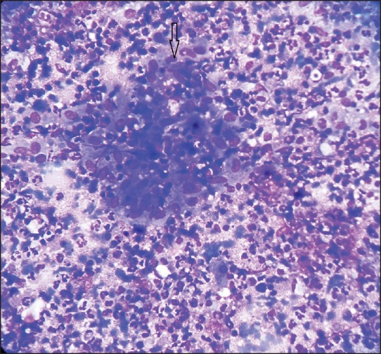
FNAC smear showing epithelioid cell granuloma (marked at arrow) in the background of inflammatory cells in case 1. (Giemsa stain, ×400)

FNAC smear showing epithelioid cell granuloma (marked at arrow) in the background of inflammatory cells in case 1. (Giemsa stain, ×400)