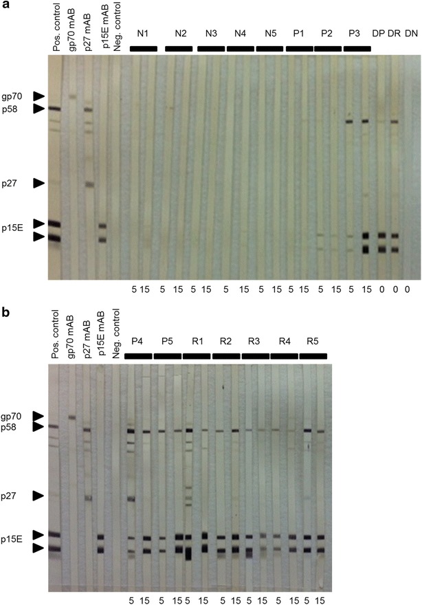
Fig. 4.

Western blot analysis using plasma samples from week 5 and 15 weeks post transfusion. The first five strips were incubated with a positive control (pool of immune cats), monoclonal antibodies against gp70, p27 and p15E, respectively, and a negative control (pool FeLV-negative cats) to characterize the respective viral proteins. a Cats N1 to N5, P1 to P3 and DP, DR and DN as indicated. b Cats P4 and P5 and R1 to R5 as indicated. Right-pointing triangle denotes bands with expected length. All samples were tested under the same assay conditions
