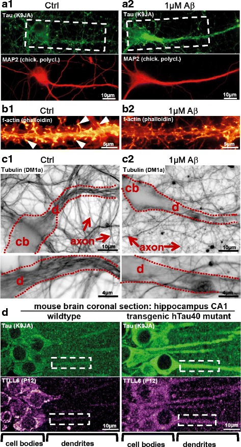
Fig. 1.

Aβ oligomers induce missorting of Tau into dendrites. Missorting of Tau correlates with dendritic loss of microtubules and mislocalization of TTLL6. a, b Micrographs of fluorescently stained primary hippocampal neurons aged for 21 days in vitro after treatment with Aβ oligomers. (a) Tau is stained with an antibody against total Tau (K9JA, green color). Staining of the dendritic marker MAP2 is shown in red. In control conditions, Tau and MAP2 do not colocalize (a1, boxed area shows a dendrite and cell body without Tau). After exposure to Aβ, Tau becomes missorted into the somatodendritic compartment, where it then colocalizes with MAP2 (a2, boxed area shows a dendrite and cell body with Tau missorting). (b) Staining of f-actin in dendritic segments with phalloidin. In control conditions, there are numerous spines (b1, f-actin positive dendritic protrusions, arrowheads). After exposure to Aβ, mature spines are lost (b2). c Microtubules were stained using an antibody against tubulin after fixing and extracting to favor microtubules over free tubulin. The filamentous structures within the dendrite in c1 (framed by dotted lines) are microtubules. In control conditions dendrites display dense microtubules (c1), whereas microtubules are lost after Aβ treatment (c2). Proximal parts of dendritic segments are magnified in lower panels. cb: cell body; d: dendrites. d Brain sections from Tau transgenic mice where missorting of Tau is present, TTLL6 is mislocalized to dendrites even without exposure to Aβ. Boxed areas highlight examples of dendritic segments. Adapted from [78]
